Ultrafast Laser Laboratory

About the Laboratory

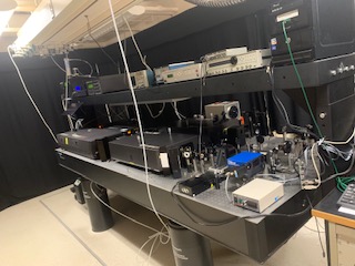
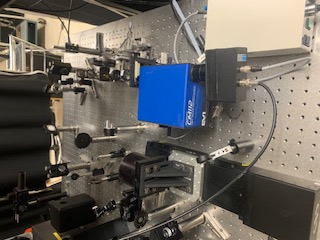
The Saskatchewan Structural Sciences Centre's Laser Technology Laboratory was built to conduct research using ultrashort pulses from Ti-doped sapphire lasers and laser amplifiers. These devices were invented in the early 1980s and since then these sources of high power short duration laser pulses have been applied for fundamental research in physics, chemistry, biology, and engineering. The short and powerful laser pulses are particularly useful for studying of compounds with very short fluorescence lifetimes. This type of laser pulses give rise to non-linear optical phenomenon in the samples under investigation, which in their turn allow to optimize the sample imaging, and give access to new contrast mechanisms for microscopy imaging. Disciplines from life sciences, chemistry, physics, and engineering make use of this lab to achieve their research goals.

Laboratory Contact

Location:
B9A
B9B

Laboratory Managers:
George Belev


Instruments and Techniques Used In Lab

This system is used as a light source for the Up-Conversion Fluorescence Lifetime instrument.

This system is used as a light source for the Time Correlated Single Photon Counting (TCSPC) instrument and as a source for the Laser Scanning Confocal Microscope. It is used for two-photon excitation imaging, fluorescence lifetime measurements (TCSPC) and for Fluorescent Life Time Imaging microscopy (FLIM).

For more detailed information, click  here

Time-Correlated Single Photon Counting (TCSPC) technique is used to measure the fluorescence lifetime of compounds in specific environments. An ultra-fast laser pulse excites the sample and the light emitted from the sample is tagged for arrival time. A decay trace of the fluorescence as a function of time is used to determine the number of lifetime components present and their respective value. Lifetime measurements from 50 picoseconds to hundreds of nanoseconds can be measured.

The SSSC fluorescence up-conversion system is used to measure the lifetime of short-lived fluorescent molecules in solution. The sample solution is exposed to an ultra-short laser pulse (400 nm) and the fluorescence emitted is monitored for intensity my mixing (up-conversion) it in a nonlinear crystal with another ultra-short laser pulse (800 nm), the probe. By delaying the time at which the probe pulse reaches the non-linear crystal, and mixes with the fluorescence, the intensity of the up-conversion can be monitored as a function of decay time. The up-conversion process is the sum frequency generated in the non-linear crystal by the combination of the fluorescence and the 800 nm probe light resulting in a wavelength shorter than that of the fluorescence. Lifetimes in the sub-nanosecond range can be measured using this technique.

Fluorescence Lifetime Imaging (FLIM) is the measurement of the lifetime of a fluorophore in an excited electronic state as a function of position on an image. This gives a picture of environment of the fluorophore. The lifetime of a fluorophore can change with chemical composition (pH, calcium concentration) or because of quenching (FRET, oxygen). At the SSSC, FLIM is based on the time domain acquisition of information in conjunction with the laser scanning confocal microscope. The pulsed laser system can used in pico- or femtosecond mode with a variety of repetition rates, and it can be used in the visible or infrared regions.

Two-photon excitation microscopy is best at reducing out-of-focus excitation, reducing the risk of bleaching a volume of the sample before the imaging process is completed. The two-photon process requires high photon density, which is achievable at the focus point of the objective with ultra-short light pulses. TPEM may reduce photo-toxicity and help to improve live cell imaging. The longer wavelength range required for the technique can make it possible to image thicker samples compared to excitation in the ultraviolet and visible ranges.

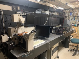
The LSCM is primarily used for imaging fluorescent samples. Organelles and proteins in cells or tissues are imaged using native fluorescence or by adding fluorescent tags. The LSCM is based on point scanning approach to image formation whereby the information for each pixel is obtained sequentially instead of the broad area excitation approach used for wide-field imaging. A laser is used for selective excitation of a fluorophore and an optical bandpass filter limits the light reaching the detector. The LSCM is useful for three-dimensional reconstruction of data. Spatial resolution ~ 300 nm is achievable with objectives of numerical aperture of 1.3.

This is a new instrument at the SSSC! Instrument details are coming soon.

Safety Information

Before coming into the lab, all users must complete “Laser Safety Awareness” course offered by the Radiation Safety Institute of Canada, found here. Upon arrival they must complete the Laboratory Specific Orientation with the research officer responsible for the Lab.

Training Information

For information regarding usage and training of the instruments in this lab, please contact the laboratory manager.