Vibrational Spectroscopy Laboratory
About the Laboratory
Vibrational spectroscopies are techniques that measure bond vibrations and allow researchers to identify various functional groups and molecular bonds within their samples. The two techniques available in the SSSC are Raman and Fourier Transform Infrared (FTIR) spectrocopies which detect changes in a chemical bond’s polarizability (ie. electron cloud around bond) and dipole moment, respectively. Both techniques are very complimentary, and can be used in many multi-disciplinary applications including:- Materials characterization
- Catalysis characterization
- Biological samples (protein secondary structure) and tissues
- Geological samples - mineral identification
- Plant materials and biomass samples
- In situ monitoring
Raman and FTIR
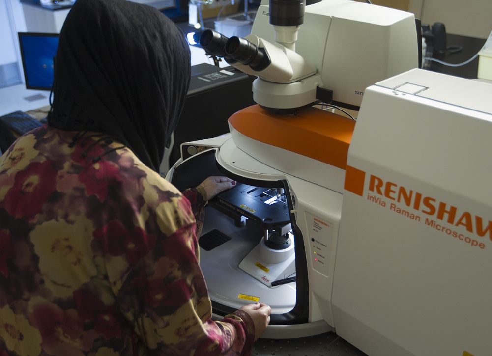
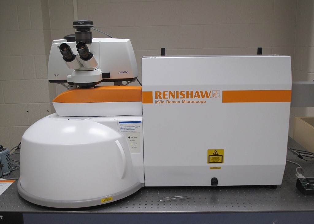
Raman and Fourier Transform Infrared (FTIR) are vibrational spectroscopies that detect changes in a chemical bond’s polarisability (ie. The electron cloud around bond) and dipole moment, respectively. Both techniques are complimentary, and many vibrational modes are both Raman and IR active. In some cases, when there is a center of inversion in the crystal symmetry of the material, some vibrational bands will be only Raman active or IR active (not both – rule of mutual exclusivity). The Renishaw Invia Reflex Raman microscope (Renishaw, Gloucestershire, UK) located within the SSSC is its only kind in Canada, as it also equipped with an IlluminatIR II FTIR microscope accessory (Smith’s Detection, Danbury, CT). In addition, the Invia Reflex microscope supports multiple laser wavelengths, along with sophisticated software that allows automatic switching of lasers. This allows multiple excitation wavelength Raman and FTIR spectroscopies to be obtained on the same spot with relative ease for the general user!
Raman or FTIR…..Which one should I use?
This is a common question we often get asked, and the choice of using Raman or FTIR spectroscopy will depend on what kind of desired information and the sample composition. Both techniques are complimentary and provide information from molecular bond vibrations. The table below describes some of the differences:
FTIR spectroscopy measures changes in the dipole moment of a chemical bond. It is well suited for identifying functional groups for organic materials, and fluorescence material is not a problem. The diamond-ATR objective is well suited for powders and other solid materials. The back-aperture can go as small as 10 µm spot size. The ARO objective is well suited for small particles and thin sections. Some of the sample limitations for FTIR include:
- Wet materials – H2O is a major contaminant in FTIR spectroscopy
- Thermal degradation – organic materials that have been degraded to amorphous carb on do not provide a good FTIR spectrum
Raman spectroscopy is a light scattering process in which inelastically scattered photons are measured from a high powered laser focussed on a sample. Raman spectroscopy measures the vibrational and lattice vibrations when there are changes in the polarizability of a molecule (ie. A distortion of the molecule’s electron cloud during vibration must cause the molecule to interact differently with the electric field of the incoming photon). Raman spectroscopy is ideally suited for:
- Graphitic materials (C=C stretch not seen in IR spectrum, but large change in polarizability)
- Various minerals and metal oxides
- Pharmaceutical products
- Polymorphisms, crystallinity distortions
- Raman confocal microscopes ideally suited for measuring embedded materials
- Raman maps with sub-mm resolution
- No interference from water – live cell imaging possible
Some of the limitations include:
Sample fluorescence.
Low S/NThe S/N can be low for some materials.

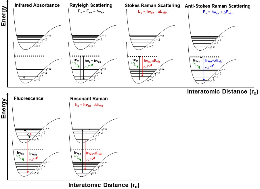
Figure 1. Infrared spectroscopy measures vibrational transitions by changes in the dipole moment of the different molecular bonds. Raman spectroscopy typically uses a high powered laser source (hνex) which excites electrons to a “virtual state” located between the electronic ground state and excited state. The majority of the photons will scatter elastically on the sample surface, and is commonly known as Rayleigh scattering. A small portion of the photons will be scattered inelastically (approx.1 part in 10 million) due to its interaction with the vibrations in the material. The Stokes Raman scattered photons are slightly absorbed by the material and will be at a slightly lower energy (red-shifted) compared to the incident photons. The antistokes Raman scattered photons gain a slight amount of energy from the material (blue-shifted). Resonant Raman spectroscopy is observed when the hνex source is near the same energy as an electronic transition. Electrons are excited into the higher electronic state and leads to an increase in Raman intensity (as much as a factor of 106). Fluorescence is the main interference of Raman spectroscopy, and generally occurs when the hνex source is near the electronic transition energy, or it could originate from the sample matrix. Because fluorescence is much longer lived excited state lifetime (τ) compared to Raman transitions, the fluorescence emission will generally mask the Raman peaks.
Instruments In Lab
The PicoRaman M2 is the second generation of the first commercially available time-gated Raman spectrometer. The use of a time-gated detector synchronized with a pulsed laser can drastically reduce the fluorescence background for some samples. Time-gating also reduces problems with ambient light or incandescence from hot samples. A variety of configuration options are available including a microscope with a motorized stage, a high temperature cell, and a probe for oversized samples.
Instrument Specifications
| Laser | Laser | 532 nm diode laser |
| Laser Power | ≤ 200 mW at laser port | |
| Pulse Energy | ≤1 µJ | |
| Pulse Repetition Rate | 100 – 250 kHz | |
| Pulse Width | < 150 ps | |
| Detector | Detector | CMOS SPAD array |
| Spectrometer Range | 0 – 2500 cm-1 | |
| Spectral Resolution | 5 cm-1, can be improved using hypersampling (grating shift feature) | |
| Time Resolution | 100 ps | |
| Gate delay range | 0 – 10 ns | |
| Microscope | Olympus BX53 with 2X (NA=0.08), 4X (NA=0.13), 10x (NA=0.3), 20x LWD (NA=0.4) and 50x LWD (NA=0.5) objectives. High temperature cell available (Linkam CCR1000, up to 1000°C) | |
The Renishaw Invia Reflex Microscope is equipped with an IlluminatIRII FTIR microscope accessory (Smith’s Detection, Danbury, CT), and it is the only one available in Canada. The microscope is equipped with a MS20 encoded stage that allows precise steps (100 nm) for high resolution mapping capabilities. The Invia Reflex has 514.5 nm and 785 nm laser excitations, along with sophisticated software that allows automatic switching of lasers. This allows multiple excitation wavelength Raman and FTIR spectroscopies to be obtained on the same spot with relative ease for the general user.
Instrument Specifications
|
Laser Pathway |
Light Path |
Filter Set |
Polarizers |
Grating (line/mm) |
|
514 nm Ar+ Laser (Modulaser StellarPro-50) |
Edge Filters (100 cm-1) |
z(x,x)z’, z(x,y)z’ |
1200, 1800, 2400 (special request) |
|
|
785 nm Laser Diode (Renishaw Inc.) |
Edge Filters (100 cm-1) |
z(x,x)z’, z(x,y)z’ |
1200 |
|
|
Light Microscope |
Leica DC2500M equipped with 5X (NA=0.12), 10X (NA=0.25), 20X (NA=0.40), 50X (NA=0.75), 100X (NA=0.85), 50X LWD (NA=0.40) |
|||
|
XYZ Motorized Stage |
MS20 Encoded Stage, Renishaw, UK) with 100 nm step sizes which allows for the capability of very high spatial resolution mapping. Coupled with the correct laser excitation and objective, and spatial resolution can be better than 0.5 µm!
|
|||
|
FTIR |
Smith’s Detection IlluminatIR FTIR microscope accessory is equipped with a liquid nitrogen cooled MCT detector allows measurements in the 4000-650 cm-1 range with up to 4 cm-1 spectral resolution. The system uses a 15X (NA= 0.88) ARO objective (10-100 µm aperture) or a 36X diamond ATR objective (10-50 µm aperture). |
|||
A recent addition to the SSSC, this Renishaw Invia Reflex Microscope is equipped with an external fiber-optic probe (λex = 514.5 nm) that allows Raman measurements on larger samples (ex. In-situ process monitoring). The microscope is equipped with a MS10 encoded stage that allows precise steps (100 nm) for high resolution mapping capabilities. The Invia Reflex has 514.5 nm and 785 nm laser excitations, along with sophisticated software that allows automatic switching of lasers.
Instrument Specifications
|
Laser Pathway |
Light Path |
Filter Set |
Polarizers |
Grating (line/mm) |
|
514 nm Ar+ Laser (Modulaser Stellar-Ren-50) |
Edge Filters (100 cm-1) |
z(x,x)z’, z(x,y)z’ |
1200, 1800 |
|
|
785 nm Laser Diode (Renishaw Inc.) |
Edge Filters (100 cm-1) |
z(x,x)z’, z(x,y)z’ |
1200 |
|
|
Light Microscope |
Leica DC2500M equipped with 5X (NA=0.12), 10X (NA=0.25), 20X (NA=0.40), 50X (NA=0.75), 100X (NA=0.85) |
|||
|
XYZ Motorized Stage |
MS10 Encoded Stage, Renishaw, UK) with 100 nm step sizes which allows for the capability of very high spatial resolution mapping. Coupled with the correct laser excitation and objective, and spatial resolution can be better than 0.5 µm!
|
|||
The SSSC has 2 portable Raman spectrometers from Enwave Optronics. The portable fiber-optic Raman spectrometers that are reliable and compact. The compact spectrometers have a λex = 532 nm or λex = 785 nm. The light-source is delivered to the sample by a high performance fiber-optic probe, allowing the instrument to be used in the lab, or taken into the field.
Instrument Specifications
|
GASRaman NOCH-2 Spectrometer (λex = 532 nm) |
|
|
Laser Excitation |
532 nm diode laser, Laser Power 0-50 mW |
|
Spectrometer Range (cm-1) |
250-4200 |
|
Spectral Resolution |
Approx. 6 cm-1 |
|
Fiber Optic Probe |
HRP-8 High throughput fiber probe sample interface |
|
Detector |
CCD camera, -60 oC |
|
Operating Temperature |
10oC to 40oC |
The SSSC has 2 portable Raman spectrometers from Enwave Optronics. The portable fiber-optic Raman spectrometers that are reliable and compact. The compact spectrometers have a λex = 532 nm or λex = 785 nm. The light-source is delivered to the sample by a high performance fiber-optic probe, allowing the instrument to be used in the lab, or taken into the field.
Instrument Specifications
|
EZRaman-I Spectrometer (λex = 785 nm) |
|
|
Laser Excitation |
785 nm diode laser, Laser Power 0-350 mW |
|
Spectrometer Range (cm-1) |
100-3300 |
|
Spectral Resolution |
Approx. 7 cm-1 |
|
Fiber Optic Probe |
High performance fiber optic probe, OD>8 |
|
Detector |
CCD camera, -50 oC |
Training Information
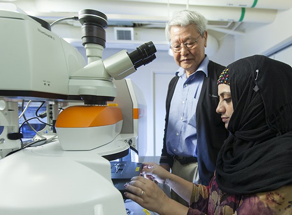
The Raman/FTIR microscope open to any academic research group. SSSC personnel will first train researchers on the instrument and offer assistance in experimental development if needed. Training typically lasts 1-2 hours. After training, researchers may use the instrument independently and reserve the instrument time through SSSC Evolution site.
Data Analysis Resources
Renishaw Wire (available at SSSC)


